


1
2
3
4
5
6
7

Confocal image of astrocytes in 1mm cleared brain slice
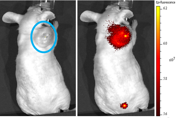
Mice bearing A549 xenografts on the right shoulder (dashed blue ovals) were injected with dye labeled vesicles derived from RAW264.7 cells demonstrating RAW vesicles specifically targeted the A549 xenograft.
Confocal imaging of vesicle delivery
Confocal image of HEK 293 cells after the delivery of vesicles loaded with fluorescein (interior) and DiD (cell membrane). The interior of the cell is filled with fluorescein and vesicles can be seen on the cell surface.
Cerebral Blood Vessels
A representative image of a 3-D reconstruction of cerebral blood vessels from in vivo 2P imaging.
Astrocytes
Cartoon art of a smoking mouse
Cover art of "Brain region specific single-molecule fluorescence imaging"